United Network for Organ Sharing
AR Organ Scanning
- Client

- Sector
- Healthcare
- What we did
- Discovery, UX design, augmented reality development, and a web viewer
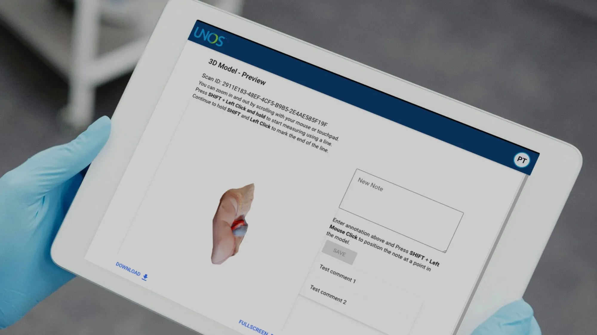
Organ evaluation, measured and shared.
The challenge
The United Network for Organ Sharing (UNOS) manages the national transplant system, and its UNOS Labs team explores new technology to improve outcomes. Organ procurement is one of the most time-sensitive processes in medicine.
Procurement teams have to scan, measure and evaluate an organ before transplant, under pressure to stay accurate and protect its viability. The tools were mostly manual, so measurements were not consistent, scans were hard to annotate and share across distributed teams, and capturing data in the field was slow. UNOS wanted to know whether augmented reality and 3D scanning could make the process more precise and more reliable.
The build
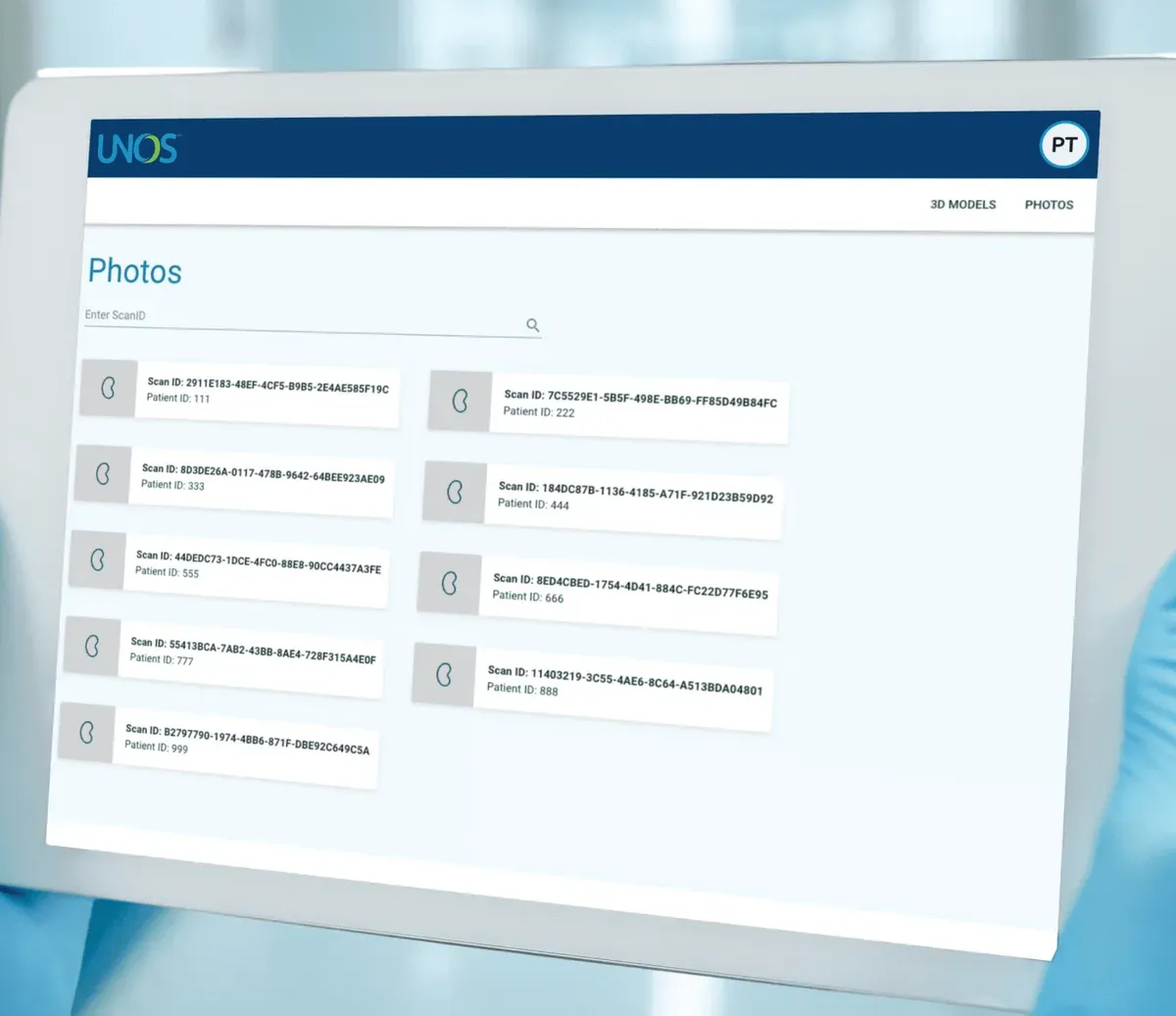
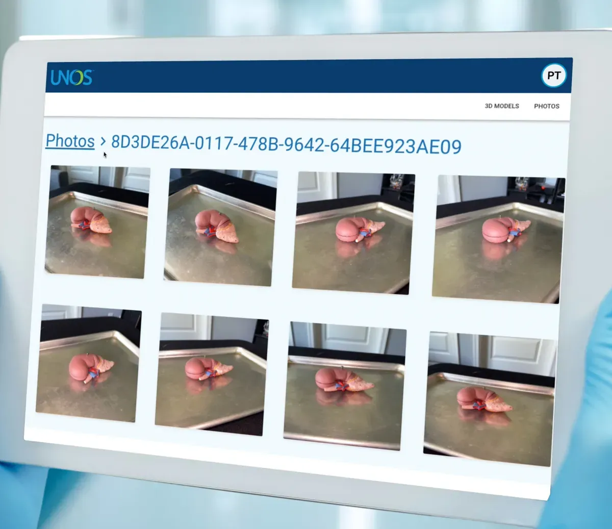
UNOS Labs asked Shockoe for an augmented reality tool, and the work ran in two phases so UNOS could test feasibility before expanding. Phase one scanned an organ in 2D, anchored the scan to scale so the organ, its arteries and its veins could be measured, recorded annotations, and rendered the result in a web viewer.
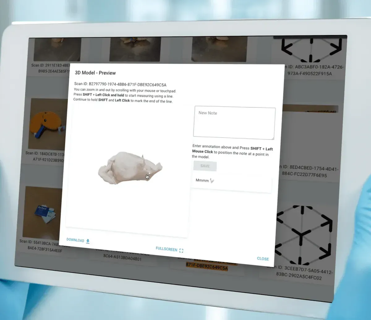
Phase two added 3D organ reconstruction, image segmentation, and better collaboration in the web viewer. The tool moved from a proof of concept to a fully deployed solution.
The result
The tool proved the approach and set the stage for digital transformation in transplant logistics. It is designed to give standardized measurements that reduce variability and support better matching decisions, to let teams evaluate organs together through shared scans and annotations, and to give UNOS a foundation to scale with 3D reconstruction.
Project images
Questions about this project
What does the AR organ scanning tool do?
It lets procurement teams scan an organ with augmented reality, measure its size and the size of its arteries and veins, and record annotations. The scans open in a web viewer, so teams can review and discuss them together.
Why did UNOS need it?
Procurement teams have to scan, measure and evaluate an organ before transplant, under time pressure and with its viability at stake. The tools were mostly manual, so measurements varied and scans were hard to annotate and share across distributed teams.
How was the work phased?
In two phases. The first phase covered 2D scanning, anchored measurements, annotations and the web viewer. The second added 3D organ reconstruction, image segmentation and better collaboration in the viewer. Both phases were delivered and the tool is fully deployed.
What can the web viewer do?
It shows the photos and 3D model captured for each scan, and lets a reviewer add notes and mark positions on the model. That keeps the scan, its measurements and the discussion in one place.

7 Ways to Drive Growth and Stay Competitive with Data and AI
Seven priorities for marketing teams using data and AI, backed by Salesforce and Gartner survey data on trust, personalization, and readiness.
Can You Explain the Code Your AI Wrote?
AI writes more code than anyone can retype. What still builds understanding is explaining it, testing it and owning what ships.
What AI Implementation Looks Like in a Large Organization With Legacy Systems
A practical guide for CIOs and CTOs: phased rollout, governance, training and integration that adds AI without replacing the systems you run on.